
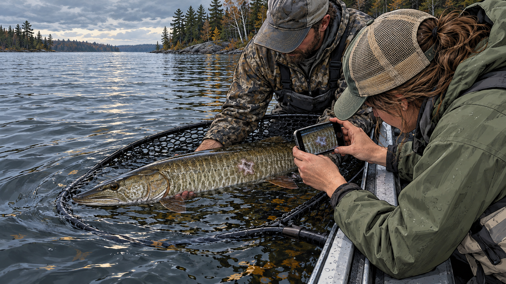
The photograph can be accurate and the diagnosis wrong
A pale patch, raised plaque, red sore, missing scale, or swollen area on a musky is an observation. Calling it fungus, cancer, a virus, hook damage, or a parasite is a diagnosis. Those are not the same act.
The distinction matters because fish-health conditions can resemble one another from above the water. The angle of the photograph, glare, mucus, old injuries, spawning abrasion, and ordinary color variation can further change what an angler thinks is visible.
The useful first move is simple: preserve the appearance and context without forcing a name onto it. The report should say what the mark looked like, where it was on the fish, and when and where the fish was encountered.
Muskies have more than one kind of skin story
Wisconsin DNR describes lymphosarcoma in northern pike and muskellunge as cancerous sores in the skin and muscle. Early signs may appear as purple or pink blisters before developing into a bulging tumor. The agency lists the pathogen as unknown and likely viral.
A pathology review describes esocid lymphosarcoma as irregular, pale-tan plaques that can merge and ulcerate. The same review documents smooth-type epidermal hyperplasia in muskellunge and northern pike as slightly raised translucent plaques that may appear blue, dark, or red. It notes that this condition must be distinguished from a grossly similar blue-spot disease.
That is the reason a color label cannot close the case. Even trained researchers use tissue structure and molecular evidence to separate conditions that can overlap in outward appearance.
Some evidence is smaller than a camera can see
During fish-health assessments in Michigan, researchers observed tiny cyst-like structures on muskellunge gill clips. They described a new myxozoan parasite only after microscopy, histology, ribosomal gene sequencing, and phylogenetic analysis. The organism was named Henneguya michiganensis.
That study did not create a field guide for diagnosing every white point on a gill. It demonstrated the opposite. The identification depended on spores, tissue response, measurements, and DNA evidence unavailable in a boat.
A camera can document a visible condition. It cannot reveal every internal lesion, identify a microscopic organism, or establish causation by itself.
A normal-looking fish can still complicate the story
A hatchery investigation isolated fathead minnow nidovirus from juvenile muskellunge experiencing lingering mortality at Wisconsin's Wild Rose Hatchery. Researchers also isolated a related strain from apparently healthy juvenile muskellunge at Michigan's Wolf Lake State Fish Hatchery.
Laboratory challenge trials then showed that the Michigan isolate was pathogenic to the juvenile muskellunge used in the experiment. The study depended on tissue examination, virus isolation, PCR, sequencing, and a controlled infection trial, not appearance alone.
The finding does not mean a healthy-looking wild adult is infected, nor does it turn every lesion into that virus. It shows why both directions of visual certainty can fail: a mark does not prove a particular disease, and an ordinary exterior does not prove the absence of a pathogen.
Build the record while the fish stays wet
Fish care still comes first. Keep the musky supported in a large rubber net in the water, prepare the camera before lifting anything, and do not extend air exposure to conduct a photo session. A report that costs the fish unnecessary handling time defeats its purpose.
When it can be done promptly, take one photograph of the entire side of the fish and one closer view of the unusual area. Include a known size reference without pressing an object into the lesion. Record the side of the body and its approximate position relative to the head, dorsal fin, lateral line, vent, and tail.
Describe only visible properties: flat or raised, isolated or multiple, intact or ulcerated, approximate dimensions, color, margin shape, and whether the gills, eyes, fins, or behavior also looked unusual. Do not scrape, swab, cut, or treat the fish.
The report needs the lake more than a dramatic caption
Wisconsin DNR asks for the waterbody, date, fish species, and approximate number of dead or dying fish. Those fields turn an isolated image into an event that a fisheries professional can evaluate against other observations.
Add the capture location at an appropriate level of precision, estimated or measured fish length, surface water temperature if actually measured, fish behavior, other affected fish observed, and the original unedited photographs. Note whether the fish swam away strongly, remained impaired, or was found dead. Do not convert those release observations into a disease conclusion.
For a fish kill, do not collect samples and do not transport fish to a DNR office or hatchery. Contact the agency first and follow its instructions. A health report never overrides current possession, harvest, size, transport, or invasive-species rules.
One fish is a case, not a prevalence estimate
One affected musky can justify a report. It cannot establish how common the condition is in the lake. Anglers do not sample fish randomly, capture probability varies, photographs are taken selectively, and the same individual can be caught more than once.
A cluster becomes more informative when observations are independently documented across dates, areas, fish sizes, and observers. Even then, professional examination is needed to determine whether the cases share a cause.
Do not count social-media photographs as separate fish unless natural markings, tags, dates, and locations make the identities defensible. A dramatic album can contain duplicates, unrelated lesions, or images from different waters.
Test a ledger, not a diagnosis
The lesion-ledger theory predicts that standardized observations will reveal patterns hidden by isolated photographs. The same lesion class may cluster by season, spawning period, waterbody, body location, size class, or the presence of a broader fish kill.
The theory gains support if independently reported observations recur in a coherent place and time and professional examination confirms that at least some share a cause. It loses support if apparent clusters disappear after duplicate fish, inconsistent descriptions, photography artifacts, and unrelated injuries are removed.
The best outcome is not an angler becoming a pathologist. It is a cleaner chain of evidence from the water to the people equipped to decide whether the observation is ordinary, isolated, or worth investigation.